ROM Paleohistology Lab
In 2009, the Vertebrate Paleontology Section at the Royal Ontario Museum installed a new rare-sample thin sectioning and paleohistology lab sponsored by an NSERC RTI grant to a cluster of researchers (PI: D. Evans, University of Toronto). This state-of-the-art research equipment is paired with the traditional paleontological resources at the Royal Ontario Museum that include a large well-equipped preparation and molding/casting lab.

Ornithomimid dinosaur bone microstructure.
The lab is open to researchers from across the country. It is intended to facilitate research on rare and valuable specimens (fossils, meteorites) that require secure curatorial housing, minimal loss of precious material, and precise execution in order to maximize research output, and avoid the risk of putting rare specimens in the hands of commercial thin-sectioning labs. The lab is operates on a cost recovery basis. To arrange access, contact Dr. David Evans.

The thin-sectioning (left) and microscopy (right) facilities at the Royal Ontario Museum.
LAB EQUIPMENT
Thin sectioning system. Equiptment includes 3 saws (including a 8” Buehler IsoMet 1000), two CrystalMaster 12″ lap wheels, and an 8” Hillquist Thin Section Machine. The large format of the saws and lap and polishing wheels maximizes the size of thin section that can be produced, including the mounting of large dinosaur bones. This set-up offers a very wide range of thin section sizes (up to 150mm by 150mm) and as such maximizes the scope of research.
Microscopy and Visualization Lab. The visualization lab includes two new microscope systems, a fully automated Nikon (AZ100 Microscope system) and an E200 Polarizing Petrographic microscope. Paired with a powerful computer, both systems uses the same Nikon imaging software. The lab is also equipped with a NextEngine desktop laser scanner and a photostand for photogrammetry using PhotoScan software.
Diamond Wire Saw Lab. Two ultra-low loss diamond wire saws were funded by an NSERC RTI grant in 2008 (NSERC # EQPEQ 359875-08, PI Ed Spooner, University of Toronto), and the facility was successfully installed in the Earth and Space Section at the Royal Ontario Museum in the Fall of 2008. Wire saws are ideally suited for cutting rare specimens, such as fossils and meteorites, because the very fine gauge of the wire blades minimizes loss of the original material, as well as a high degree of precision in taking the rough sample prior to embedding and mounting on a slide. This equipment is complementary to the thin section facility here, and together these two infrastructure initiatives at the ROM form a highly integrated, world-class facility for rare sample research.
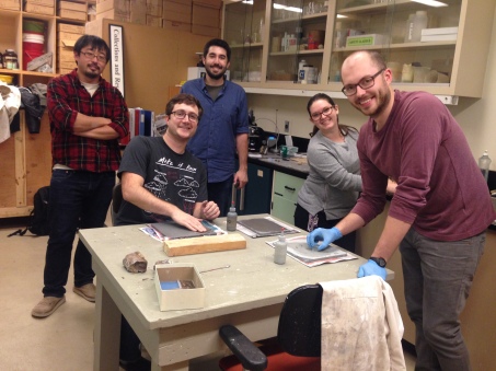
University of Toronto grad students using the lab.
PUBLICATIONS
- Brink, K.S., Reisz, R.R., LeBlanc, A.R.H., Chang, R.S., Lee, Y.C., Chiang, C.C., Huang, T., and Evans, D.C. 2015. Developmental and evolutionary novelty in the serrated teeth of theropod dinosaurs. Scientific Reports 5: 12338 DOI: 10.1038/srep12338.
- Cullen, T. M., D. C. Evans, M. J. Ryan, P. J. Currie and Y. Kobayashi. 2014. Osteohistological variation in growth marks and osteocyte lacunar density in a theropod dinosaur (Coelurosauria: Ornithomimidae). BMC Evolutionary Biology 14(1):231. Available here.
- Evans, D. C., P. M. Barrett, K. Brink, and M. Carrano. 2014. Osteology and bone microstructure of new, small theropod dinosaur material from the early Late Cretaceous of Morocco. Gondwana Research. DOI: 10.1016/j.gr.2014.03.01
- Brink, K. S. and R. R. Resiz, D. 2014. Hidden dental diversity in the oldest terrestrial apex predator Dimetrodon. Nature Communications. doi: 10.1038/ncomms4269
-
Song, W, TW de Haas, H Fadaei, D Sinton. 2014. Chip-off-the-old-rock: the study of reservoir-relevant geological processes with real-rock micromodelsLab on a Chip. Advance access here.
-
Reisz, R. R., Huang, T. D., Roberts, E. M., Peng, S., Sullivan, C., Stein, K., … & Zhong, S. (2013). Embryology of Early Jurassic dinosaur from China with evidence of preserved organic remains. Nature, 496(7444), 210-214.
- Brink, K. S., LeBlanc, A. R., & Reisz, R. R. (2014). First record of plicidentine in Synapsida and patterns of tooth root shape change in Early Permian sphenacodontians. Naturwissenschaften, 1-10.
-
MacDougall, M. J., LeBlanc, A. R., & Reisz, R. R. (2014). Plicidentine in the Early Permian Parareptile Colobomycter pholeter, and Its Phylogenetic and Functional Significance among Coeval Members of the Clade. PloS one, 9(5), e96559.
- LeBlanc ARH, Reisz RR (2013) Periodontal Ligament, Cementum, and Alveolar Bone in the Oldest Herbivorous Tetrapods, and Their Evolutionary Significance. PLoS ONE 8(9): e74697. doi:10.1371/journal.pone.0074697
ROM PALEOHISTOLOGY WORKSHOPS
For more information on the annual ROM paleohstology workshop, visit this page.
